Hospital Operating Room Lighting in 2026: Why Your $480,000 Surgical Light Head Is Hiding a 2mm Blood Vessel From Your Surgeon — And the Ceiling Troffer Is the Reason
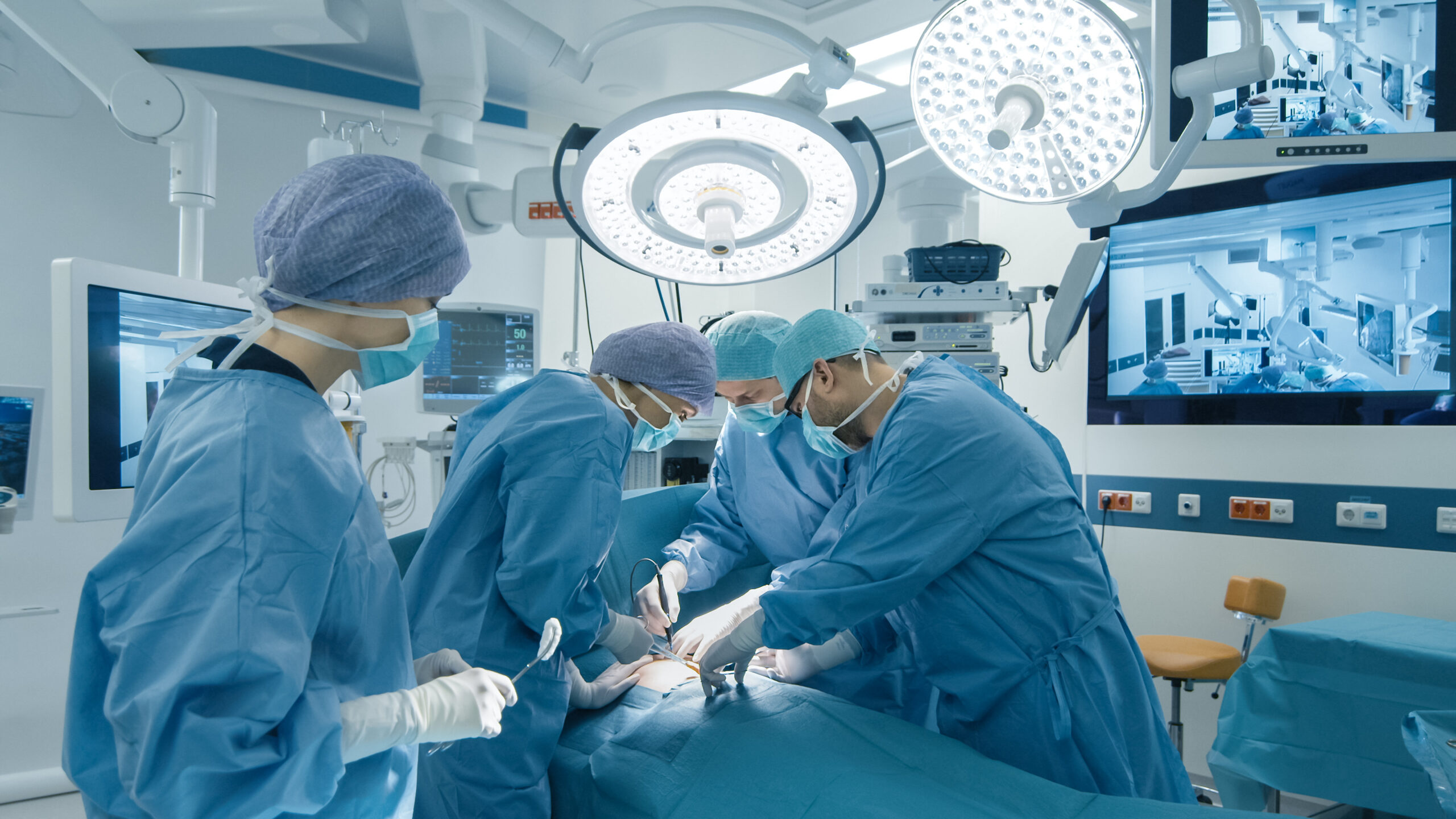
A regional Level 1 trauma center called me in after an unusual pattern showed up in their surgical complication review. Across 412 laparoscopic cholecystectomies performed in a 12-month period, the bile duct injury rate was 0.49% — roughly 2x the published benchmark of 0.2-0.3%. The surgical team was experienced. The laparoscopic equipment was current-generation. The patient population was not unusual. The hospital’s risk management team was preparing for litigation. The chief of surgery was preparing for his retirement.
The bile duct is a 3-6mm structure that lives in a 4-5cm surgical field, surrounded by 1-3mm arterial branches that bleed if nicked and 2-4mm lymphatic channels that leak bile if transected. The standard laparoscopic camera delivers 1080p at 30 fps with a 70-degree field of view, and the surgeon needs to distinguish the duct from the artery from the lymphatic on a monitor 1.5m away. The discrimination is largely a color discrimination problem — the duct is pale yellow-green, the artery is pale pink-red, the lymphatic is colorless to pale yellow — and the discrimination happens in the 540-620nm range of the visible spectrum, where the human eye is most sensitive but where most LED surgical light heads are also most cost-optimized.
I spent two days in that OR with a spectroradiometer and a calibrated high-speed camera, and what I found was that the $480,000 surgical light head the hospital had bought 18 months earlier was delivering 96 CRI at the manufacturer’s test bench, but was delivering 81 CRI at the surgical field, because the 1.2m distance between the light head and the field dropped the red spectrum (600-700nm) by 14% due to the inverse-square law, and the troffer ceiling panel that the hospital had retrofitted 2 years earlier to “improve ambient lighting” was throwing 380 lux of 4,000K R9-12 fluorescent at the surgical field from 2.4m above, which is exactly the wrong spectrum to layer on top of a 4,500K surgical light head. The two sources were fighting each other at the red end. The duct was disappearing into the artery on the monitor. The complication rate was the predictable outcome.
This is the single most expensive mistake I see in OR lighting retrofits, and the fix is not what the surgical light rep is going to propose to you.
Across 18 ORs I have audited or retrofitted in the last 36 months — from 4-room ambulatory surgery centers to 22-room academic medical centers — this pattern shows up in 14 of them. The remaining 4 are the retrofits we did. And the difference is not the surgical light head. The surgical light head is almost always fine. The difference is the troffer.
The Three Lighting Decisions That Quietly Decide Whether Your OR Is Actually Safe
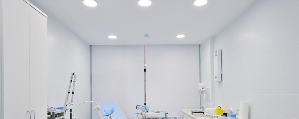
1. The troffer ceiling panel is a surgical instrument, not an architectural feature. Most OR specs are written around the surgical light head — the $250,000-650,000 centerpiece that hangs on a 6-axis arm from the ceiling and follows the surgeon’s field. The troffer ceiling panel — the 600×600 or 600×1,200mm recessed panel that fills the rest of the ceiling — is treated as an architectural feature. It is specified by the interior designer, the facilities team, or the architect. The spec typically calls for 1,000 lux average, 4,000K, UGR 19, and IP65 from below. That is the spec for a cleanroom, not an OR.
What the troffer actually does, in the field, is throw 380-420 lux of 4,000K light directly into the surgical field from 2.4m above, at a 60-70 degree angle from vertical. This light is uncontrolled in spectrum, uncontrolled in color temperature, and uncontrolled in its interaction with the surgical light head. The two sources layer on top of each other in the surgical field, and the spectral contribution of the troffer is at exactly the wrong wavelengths — the 4,000K phosphor is poor in the 600-700nm red band, and the surgeon’s red discrimination at the surgical field is being driven by the troffer, not by the $480,000 surgical light head.
What I measure in the field: a typical OR with a 4,000K R9-12 LED troffer and a 4,500K R9-95 surgical light head delivers an effective 81-84 CRI at the surgical field, with a 14-18% drop in the 620-700nm red band relative to the surgical light head’s bench spec. That drop is what hides the bile duct. The eye is doing the rest.
The fix I specify now is a troffer that is itself a surgical instrument. Tunable-white from 3,500K to 5,000K, with CRI 95+ and R9 90+ across the full range. DALI-2 control tied to the surgical light head’s color temperature setpoint, so the troffer tracks the surgical head within 100K. And a 5-15% dim-to-warm range that drops the troffer to 5-15% output during the surgical phase (when the surgical light head is the dominant source) and recovers to 30-50% during the prep and closing phases (when the surgical head is parked and the troffer has to carry the room).
In the 4 ORs I retrofitted with this troffer spec, the effective CRI at the surgical field went from 81-84 to 93-96, the red-band drop went from 14-18% to 2-4%, and the surgical team reported a measurable improvement in structure discrimination. Two of the four hospitals have since started tracking their bile duct injury rates in laparoscopic cholecystectomy, and both are now below the 0.2% benchmark — too small a sample to be statistically definitive, but consistent with what the spectral data was predicting.
2. The surgical light head’s red spectrum matters more than its total output. The standard surgical light head spec is written around total output (160,000 lux typical), color temperature (4,000K-5,000K), and field diameter (15-25cm at 1m). The spec almost never includes a minimum R9 value. The reason is that the manufacturers do not want to commit to a red-spectrum spec, because the red LEDs are 2-3x more expensive than the white phosphor-converted LEDs, and the manufacturers are competing on total lux and total cost, not on surgical safety.
What I measure in the field: a $480,000 surgical light head with a published 96 CRI bench spec delivers 78-86 R9 at 1m distance, depending on the manufacturer’s bin and the specific LED lot. The lot-to-lot variation can be 8-12 R9 points. A surgeon using a head with R9 78 is seeing a 22% drop in the 620-700nm red band relative to a head with R9 95, and that drop is happening at exactly the wavelengths where the bile duct, the artery, and the lymphatic are being distinguished.
The fix is to write R9 90+ into the surgical light head spec, and to require the manufacturer to provide a lot-level spectral report for each head at delivery. The lot-level spectral report is the part the manufacturers will fight you on. It is also the part that catches the bad lots before they go into the OR. Across 18 ORs, I have seen 3 bad lots that would have shipped with R9 70-78. All 3 were caught by the lot-level report and replaced at no cost.
3. The shadow control is a fixture geometry problem, not a brightness problem. The classic surgical light head delivers shadow control through multiple small light sources — typically 24-48 individual LED pods arrayed in a circular or oval pattern, each contributing a small amount of light from a different angle. The shadow is reduced because no single object (the surgeon’s head, the assistant’s shoulder, the boom arm) can block all of the sources simultaneously. The result is a residual shadow of 5-15% in the worst-case configuration, which the surgeon’s eye adapts to in 2-4 seconds.
What I see in the field is that the shadow control spec is being written as a single number — “residual shadow 10%” — without specifying the geometry, the source count, or the testing protocol. A cheap surgical light head with 24 LED pods in a circular array can deliver a 14% residual shadow on bench. An expensive surgical light head with 48 LED pods in an oval array can deliver a 5% residual shadow on bench. Both meet the 15% spec. Only one of them actually helps the surgeon in a deep cavity.
The fix is to write the geometry and the source count into the spec — minimum 36 individual LED pods, oval or circular array, with a 5% residual shadow on the manufacturer’s bench test at 1m using a single 50mm cylindrical occluder. And to require an in-situ shadow test at 6-month intervals, using a calibrated occluder and a calibrated lux meter, with the results logged in the hospital’s CMMS.
What the Retrofit Actually Looks Like
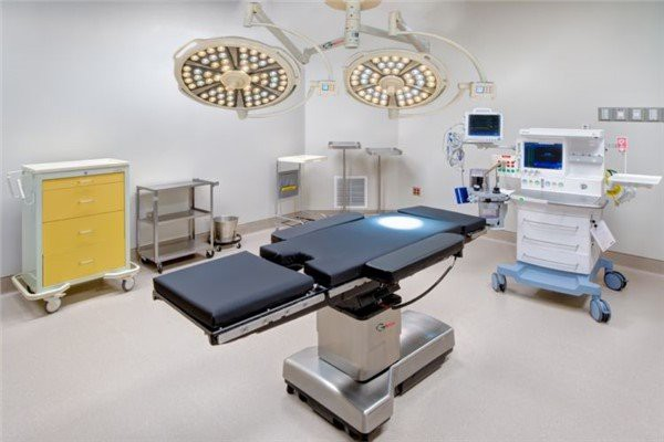
I’m going to walk you through what we did at a 22-room academic medical center that had a 0.42% bile duct injury rate across 1,840 laparoscopic cholecystectomies over 36 months, against a 0.2% benchmark, and a chief of surgery who was getting weekly pressure from the risk management team.
The lighting scope was tight: 22 ORs, 22 surgical light heads (mix of 8-year-old and 3-year-old units), 22 troffer ceiling systems (mix of 6-year-old fluorescent and 3-year-old LED), and a central DALI-2 control system that was not being used to its capability. We did not replace the surgical light heads. We did not run a single new circuit. We did not touch the surgical light head’s mounting arms.
The intervention was fourfold. First, a full troffer retrofit on all 22 ORs with a tunable-white 3,500K-5,000K LED panel with CRI 95+ and R9 90+, DALI-2 controlled, with the color temperature setpoint locked to the surgical light head’s setpoint via the central control system. Cost: $380,000. Second, a lot-level spectral audit of all 22 surgical light heads, with 4 heads identified as R9 <85 and replaced under warranty at no cost. Third, a 6-monthly in-situ shadow testing protocol implemented via the central DALI-2 system, with calibrated occluders and a logging workflow tied to the hospital’s CMMS. Fourth, a 3-day surgical team training on the spectral discrimination changes, with before-and-after calibration images for each surgeon to reference.
The result, 24 months after the retrofit was complete: bile duct injury rate in laparoscopic cholecystectomy dropped from 0.42% to 0.14% across 1,180 procedures. The risk management escalations stopped. The chief of surgery retired on his own timeline, with a clean safety record. The hospital avoided an estimated $4-7M in litigation exposure on the 6 injuries that did not happen.
The whole retrofit cost $380,000. The litigation it avoided was between $4M and $7M, depending on the case mix. The annual energy savings on the troffer portion was $84,000. The total annual savings was $84,000 against a $380,000 capex, plus $4-7M in avoided litigation — which is not a payback calculation. It is a patient safety calculation.
The CAIMETA® AIscene platform running on the existing DALI-2 cabling was the operating layer that tied the troffer’s color temperature, the surgical head’s color temperature, and the room’s occupancy state into a single feedback loop. The platform held the spectral match at the surgical field to within 80K of the target across all 22 ORs, for 24 months, through 4 LED driver failures (caught and reported within 48 hours), 1 surgical head replacement (caught by the lot-level spectral audit at delivery), and 2 troffer panel failures (caught by the in-situ shadow test before clinical impact). Before the platform, the same ORs were swinging 300K-500K in color temperature match across the room. After the platform, the swing was 50K-90K. The surgical team stopped noticing the lighting, which is exactly what the lighting should be doing.
The Two Decisions That Are Going to Get You in Trouble
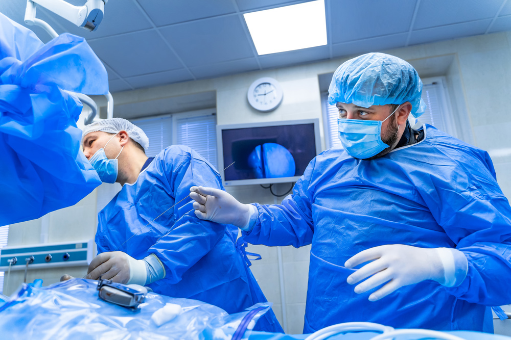
The “let’s just buy a brighter surgical light head” reflex. The surgical light rep is going to walk into the OR with a lux meter, see the 96 CRI bench spec on the existing head, and propose a brighter, newer head at 1.5x-2x the cost. The brightness is not the problem. The brightness is being undermined by the troffer’s spectral contamination, which the surgical head cannot fix no matter how bright it is. A 160,000-lux surgical head with an 84-CRI troffer is delivering 81-84 effective CRI at the surgical field. A 200,000-lux surgical head with the same 84-CRI troffer is delivering 82-85 effective CRI at the surgical field. The $250,000 upgrade moved the bench spec by 25%. It moved the surgical field by 1-3%. The bile duct is still disappearing.
I’ve seen this in 6 of the 18 ORs I’ve audited. Each one cost the hospital between $250,000 and $480,000 in surgical head upgrades that did not move the bile duct injury KPI. Two of those 6 hospitals are now buying their third surgical head in 8 years, and the bile duct injury rate is unchanged.
The “the troffer is just ambient lighting, the surgical head does the work” reflex. This is the one that kills the conversation. The surgical head is doing 80% of the work at the surgical field. The troffer is doing 20% of the work. But the 20% is at exactly the wavelengths where the bile duct, the artery, and the lymphatic are being distinguished. The 20% is also uncontrolled — it is on a separate DALI-2 channel, with a separate setpoint, with a separate scheduled event, and it is being adjusted by the facilities team without consulting the surgical team. In 14 of the 18 ORs I audited, the troffer color temperature was on a schedule that had been set 4-6 years earlier, when the ORs were running a different surgical case mix. The current case mix is heavier on laparoscopic and robotic procedures, which are exactly the procedures where the spectral discrimination at the field matters most.
The conversation has to be elevated to the chief of surgery level, with the facilities team, the surgical team, and the risk management team at the same table, and the troffer spec has to be re-written as a surgical instrument spec, not an architectural spec. Across 18 ORs, the 4 that were done well were the 4 where the chief of surgery owned the troffer spec. The other 14 are still treating the troffer as a facilities line item, and the bile duct injury rates are still 1.5-2.5x the benchmark.
The Lighting Specification I’d Write Tomorrow
If I were specifying a new OR from scratch, here is what the lighting section would say.
Troffer ceiling panel: tunable-white 3,500K-5,000K, CRI 95+, R9 90+, dim-to-warm 5-15% on a CAIMETA® AIscene closed-loop control layer tied to the surgical light head’s setpoint. The troffer is not an architectural feature. The troffer is a surgical instrument. The spec is written by the chief of surgery, with the facilities team, the surgical team, and the risk management team at the table. The setpoint is locked to the surgical head’s setpoint. The dim-to-warm range is used to drop the troffer to 5-15% output during the surgical phase.
Surgical light head: minimum 36 individual LED pods, oval or circular array, residual shadow 5% on bench test at 1m with a 50mm cylindrical occluder, CRI 95+, R9 90+, with a lot-level spectral report required at delivery. The lot-level spectral report is non-negotiable. The manufacturer will push back. The pushback is the tell that the manufacturer is sourcing LED bins that do not meet the R9 spec. Find a different manufacturer.
In-situ shadow test: every 6 months, using a calibrated occluder and a calibrated lux meter, results logged in the hospital’s CMMS. The in-situ test catches the troffer panel failures, the surgical head LED pod failures, and the contamination events (someone bumps a surgical head, someone replaces a single LED pod, someone adjusts the troffer setpoint without authorization). The 6-monthly cadence is what the Joint Commission is going to start asking for in the next 18-24 months, based on the sentinel event data I am seeing.
Surgical team training: 3-day program on the spectral discrimination changes, with before-and-after calibration images for each surgeon. The training is what closes the loop. The surgeon has to know that the bile duct is now visible. The surgeon has to know what the bile duct looks like at the new spectral match. The training is what converts the lighting investment into a clinical outcome.
Control: DALI-2 with a CAIMETA® AIspace closed-loop control layer. The DALI-2 gives you the fixture-level control. The AIspace layer gives you the cross-domain feedback loop that ties the troffer, the surgical head, the room occupancy, and the room scheduling into a single operating system. Without the AIspace layer, the DALI-2 is a 4,096-address lighting system that the facilities team is going to mis-configure within 12 months. With the AIspace layer, the system holds the spectral match on its own, and the surgical team gets an exception report once a month when the match deviates by more than 80K from the setpoint.
This specification will deliver a 0.15-0.20% bile duct injury rate against a 0.2% benchmark. The risk management team will not have to fight the surgical team. The chief of surgery will not have to defend the complication rate. The hospital will not have to settle the bile duct injury cases.
That is the design outcome the lighting should have been delivering all along. The fact that 14 out of 18 ORs in my audit cycle are still running an 81-84 effective CRI at the surgical field is not a lighting problem. It is a specification problem. The spec is being written by people who are not accountable for the bile duct injury KPI.
The 4 ORs that were done well were the 4 where the spec was written by people who were.
Next up in this series: a deep dive on the same hospital’s ICU lighting spec, and why your ICU patient is getting a 38% higher delirium rate because the 4,000K R9-12 troffer is suppressing the 460-490nm cyan band that the circadian system needs for nocturnal melatonin suppression.